Potential ‘Game-Changer’ for Treating Pancreatic Cancer

920x920

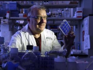
M.D. Anderson study called potential ‘game-changer’ for treating pancreatic cancer

M.D. Anderson researchers may be able to catch pancreatic tumors early

Researchers at M.D. Anderson Cancer Center believe they have found a path to early detection that could yield more effective treatment of pancreatic cancer, an illness so lethal that just 7 percent of patients survive for five years after their diagnosis.

In a study published in Wednesday’s issue of the science journal Nature, the researchers found patients with pancreatic cancer had certain particles in their bloodstream not found in healthy patients or in those with other types of pancreatic disorders. The presence of those particles could determine, with 100 percent accuracy, who had pancreatic cancer and who did not, the study found.

While the test must undergo more rigorous study before it can be used routinely, it could finally provide a reliable, noninvasive way of finding the cancer before it’s too late to treat it.

“If it pans out, it’s a game-changer,” said Dr. Raghu Kalluri, chair of cancer biology at M.D. Anderson and a co-author of the study, “and we’re aware that our patients really need that right now.”

The National Cancer Institute estimates 48,960 new pancreatic cancer cases will be diagnosed this year, and it will kill 40,560 people.

Pancreatic cancer has among the worst outcomes of any cancer. The pancreas is located deep within the body, so early tumors cannot be seen or felt, and four out of five patients are diagnosed only after it has spread. Patients usually have no symptoms until the cancer has spread to other organs. While surgery to remove the tumor or entire organ can cure up to 50 percent of early-stage pancreatic cancer, doctors see few patients in that stage.

“It’s almost invariably advanced,” said Dr. Kirk Heyne, a medical oncologist at the Houston Methodist cancer center who was not involved in the study. “Probably 15 percent of people can be taken to surgery.”

‘Unheard of’ accuracy

Even after diagnosis, there are no good ways of tracking what is happening with the cancer. Patients must undergo repeated biopsies or CT scans to determine whether the treatment is working or whether their cancer has progressed or recurred.

Researchers found the marker by looking at exosomes, tiny bundles containing DNA and other genetic material, released by the body’s internal cells.

They compared exosomes from healthy and cancerous cells and found the cancer exosomes contained a particular protein. By searching for exosomes with this protein in the bloodstream, the researchers could detect and quantify the amount of cancer in the body.

They then screened the blood of hundreds of pancreatic cancer patients, diagnosed at different stages, and hundreds of healthy donors for exosomes containing the protein. In addition to M.D. Anderson, institutions in Germany and Spain participated in the study.

“When it was positive for it, it was always in the cancer patients,” Kalluri said, “never in the hundreds of healthy subjects or those who didn’t have pancreatic cancer. The fine difference with which it was able to distinguish is unheard of in the cancer field.”

The test had zero false positive and zero false negative results.

Moreover, the study found the more advanced the patient’s cancer, the more exosomes in their blood, and when tumors were removed with surgery, levels of the exosomes dropped.

Pancreatic cancer experts called the study promising and were eager to see if further research would confirm the accuracy of the test.

“Blood-based biomarker studies for pancreatic cancer have been somewhat disappointing overall,” said Dr. Michael Goggins, director of the Pancreatic Cancer Early Detection Laboratory at Johns Hopkins University. “This one has some promising characteristics.”

The test could be used primarily in patients at higher risk for pancreatic cancer, such as those who are obese, diabetics, smokers or those with a family history of the disease.

(click here to continue reading this article)

*credit verbatim via http://www.houstonchronicle.com *


Blood Test Could Potentially Diagnose Pancreatic Cancer Early

Due to the lack of reliable early detection methods, the vast majority of pancreatic cancer cases are diagnosed once the disease has spread locally or widely throughout the body. Patients whose disease is diagnosed when it’s still confined in the pancreas have the best chance for long-term survival, which is why improved early detection strategies are urgently needed for pancreatic cancer.

A paper, entitled “Glypican-1 identifies cancer exosomes and detects early pancreatic cancer,” was published online in the prestigious journal Nature on June 24, 2015, and describes a promising, potential pancreatic cancer early detection biomarker. A biomarker is a molecular clue inside the body that can be detected and measured, providing useful information.

The paper reports that pancreatic cancer cells emit small particles called exosomes, which contain DNA and proteins. Unexpectedly, the research team from MD Anderson and abroad found that pancreatic cancer-derived exosomes contain significant levels of a protein called glypican-1 (GPC1), which is detectable at much higher levels in blood samples from pancreatic cancer patients, compared to healthy individuals. Even people with other pancreatic diseases, like chronic pancreatitis, had levels of GPC1 similar to healthy individuals, suggesting that its presence may be specific to cancer. Moreover, higher levels of GPC1-positive exosomes were found in every pancreatic cancer patient that was evaluated. By contrast, GPC1-positive exosomes were detectable in some, but not all, breast cancer samples analyzed.

In addition to blood samples from patients with advanced pancreatic cancer, the research team analyzed samples from five individuals with confirmed precancerous abnormalities in their pancreas. Encouragingly, these samples were distinguishable from healthy control individuals, suggesting that this method of detecting and measuring GPC1-positive exosomes in blood samples holds promise for the earlier detection of pancreatic cancer.

It is important to note that these results, while promising, are preliminary and have not been verified in a prospective, or forward-looking, study. More research will be necessary to determine whether the authors’ results can be confirmed as well as whether and how detection of GPC1-positive exosomes could be standardized for use in a clinical setting, rather than in a laboratory.

For more information about this article, please refer to the press release issued by the University of Texas MD Anderson Cancer Center.

For information about pancreatic cancer diagnosis, treatment and other patient services, please call 877-272-6226, Monday – Friday, 7 a.m. – 5 p.m. PT, or email a Patient Central Associate at patientcentral@pancan.org.

(click here to continue reading this article)

*credit verbatim via http://www.pancan.org *

Breast cancer surgeon, 55, dies just one month after being diagnosed w/ Pancreatic Cancer

A sad, unfortunate example of how deadly pancreatic cancer truly is:

Leading breast cancer surgeon, 55, dies just a month after being diagnosed with pancreatic cancer

298368F500000578-3118645-image-m-2_1433953709126

Tributes have poured in for one of North Carolina’s leading breast cancer surgeons who passed away from cancer at the weekend – just 33 days after she was diagnosed.

Dr. Teresa Flippo-Morton, 55, succumbed to the disease on Sunday, little over a month since she learned she had pancreatic cancer that had spread to her liver.

Former colleagues and patients – some of whom credit her with saving their lives – have paid tribute to the skilled surgeon and married mother on a Caring Bridge page she set up after her diagnosis.

She treated an estimated 2,300 women with breast cancer after completing her training in 1991, Dr. Richard White, a friend and colleague at the Levine Cancer Institute, told the Charlotte Observer.

‘This is a massive loss for our community,’ he told the newspaper. ‘She was loved by her patients. She was loved by her peers. She was loved by her students.’

The West Virginia-native, who worked at the Levine Cancer Institute, started feeling abdominal and back pain at the end of April and a scan revealed a mass on her pancreas on May 5. Further tests showed that it had spread to her liver.

She took a previously planned trip to the United Kingdom before she returned and started chemotherapy on May 21.

Following the diagnosis, she set up a Caring Bridge page to keep loved ones updated with her treatment. The page has now been visited nearly 20,000 times.

She thanked her family and friends for her support as she started her treatment.

‘I am completely moved by the cards,letters, texts, emails, and posts to this site!’ she wrote from hospital on May 20. ‘I felt immediately the warm all-encompassing love and support from everyone.’

Others took to the page to express their thanks to her.

‘You have touched the lives of so many people in so many ways as you learned and then practiced your calling,’ wrote Lee Ann McGinnis on May 31. ‘Your openness and honesty this past month is a reflection of the integrity with which you have always lived your life. You are a blessing to us.’

Another added: ‘You are the most compassionate and gracious doctor that I have ever met. You have truly touched so many lives for the better, as you have touched mine.’

On May 24, Dr. Patrick Connor, a fellow surgeon who first met Flippo 25 years ago while he was on his residency, expressed his shock and sadness at her diagnosis.

‘It is so difficult to get one’s arms around how someone so kind, caring and smart who has spent her entire professional life dedicated to the treatment and care of patients with cancer is now fighting the battle from a different perspective,’ he wrote.

(click here to continue reading this article)

*credit verbatim via http://www.dailymail.co.uk *

Treatment Doubles Progression-free Survival in some PanCan Patients

Enzyme lowers pressure inside tumors, allows chemotherapy in

Combining chemotherapy with an experimental enzyme that lowers pressure within tumors seems to double the amount of time certain patients with metastatic pancreatic cancer have before their disease progresses, suggests an interim analysis of data from an ongoing phase 2 trial. However, the trial must be completed before any definitive conclusions can be drawn, cautioned study principal investigator and pancreatic cancer expert Dr. Sunil Hingorani of Fred Hutchinson Cancer Research Center.

“For me, this ― at an absolute minimum ― says ‘finish this trial,’” said Hingorani, who is presenting the interim results of the Halozyme Therapeutics-sponsored Halo 202 trial this Sunday at the annual meeting of the American Society for Clinical Oncology. The meeting, which begins today in Chicago, runs through June 2.

The trial combines the latest first-line, standard-of-care combination chemotherapy for advanced pancreatic cancer with an enzyme called PEGPH20 that breaks down hyaluronic acid, or HA. HA is a molecule that absorbs water and serves as a natural shock absorber in knee joints. But it’s also produced in high amounts in some tumors and has been linked to a poor prognosis.

In high-HA tumors, the water-loving HA sucks the moisture from surrounding tissues into the tumor, leading to such high pressures that drugs travelling through the bloodstream can’t get in to effectively attack cancer cells.

PEGPH20 is a version of the naturally occurring hyaluronidase enzyme that breaks down HA, which has been modified to be more stable in the body. The idea behind the trial, generated by mouse model research published in 2012 by Hingorani’s team, is that using the enzyme to break down HA and lower the pressure inside the tumors will allow blood to penetrate and deliver cancer-killing drugs to their targets.

Last September, the U.S. Food and Drug Administration granted fast-track status to Halozyme’s PEGPH20 program in pancreatic cancer, a designation that is designed to speed the development and review of drugs that fill an unmet need in treating serious medical conditions.

On Sunday, Hingorani will present results from 135 participants in the trial. So far, these preliminary data show that of the patients with high-HA tumors, those who received the experimental enzyme with their chemo experienced an average of 9.2 months of progression-free survival, more than double the 4.3 months of those with high HA who were treated with chemo but no enzyme. (For the patients with low HA, the enzyme didn’t seem to make a difference; these patients had only around five months of progression-free survival, on average, regardless of whether they had received the enzyme.)

The progression-free survival conferred on the high-HA patients by the experimental treatment is higher than the overall survival (a much more definitive survival measure) seen in a recently completed phase 3 trial of the combination chemotherapy — “which is what makes this so surprising,” Hingorani said.

Halozyme Therapeutics presented these preliminary results to analysts and investors in January. Reuters reported that CEO Helen Torley said PEGPH20 is “a blockbuster in waiting” and cited the wide range of solid tumors known to produce high levels of HA. Halozyme plans to begin a phase 3 trial of PEGPH20 in patients with high-HA pancreatic tumors early next year.

Halo 202’s preliminary results hold out the prospect of doubling the remaining life spans of many patients with metastatic pancreatic cancer, a disease in which survival after diagnosis is measured in months. But Hingorani stressed that these are still small numbers of patients and the results are still preliminary. “I look at this and I say, ‘OK, the hypothesis is still intact,’” said Hingorani, who has no personal financial interest in the study and said he has never owned any of Halozyme’s stock.

But he’s hopeful.

(click here to continue reading this article)

*credit verbatim via http://www.fredhutch.org *

Tumor Molecule Discovered That Causes Spread of #PancreaticCancer

A single molecule switches on metastasis, or spread, in pancreas cancers, reports new research led by scientists at Fred Hutchinson Cancer Research Center. The researchers predict that testing for this molecule, called RUNX3, could soon help oncologists choose the most appropriate treatments based on the metastatic potential of each patient’s disease.

“We’re defining a readout that may help doctors in their approach to treatment of patients who have pancreatic cancer,” said Dr. Martin “Marty” Whittle, a postdoctoral researcher in the lab of Dr. Sunil Hingorani and first author on the paper, which was published online today in the journal Cell. “The gene that we identified can be used to give some insight as to whether a patient’s tumor is more likely to grow locally or metastasize.”

RUNX3, the researchers found, controls the activation of numerous genes involved in metastasis in a mouse model, triggering cancer cells to migrate to other parts of the body and turning on genes that help those metastatic cells take root and thrive once they invade distant tissues.

“It’s extraordinary — it seems to control an entire metastatic program,” said Hingorani, the senior researcher on the study and a physician-scientist at Fred Hutch who specializes in pancreatic cancer. “RUNX3 serves to both expel the seed and prepare the soil.”

Pancreatic cancer has the highest metastatic drive of any malignancy, Hingorani said. By the time they are diagnosed, most pancreas cancer patients already have either metastatic disease or tumors that have grown too much to be surgically removed. Even when patients’ tumors are contained, making them eligible for surgery or focused radiation, many of them end up dying of metastatic cancer anyway — because their apparently localized tumors had nevertheless already started spreading to other sites.

For this reason, oncologists often treat patients who have early-stage pancreas tumors with chemotherapy before surgery in the hopes of killing off any distant microscopic metastases and prolonging life. But in the time it takes for a few rounds of chemo, almost a third of surgically removable tumors grow too large to be eligible for surgery, a “devastating” outcome, said Hingorani, which slashes patients’ average survival time from two years for a removable tumor to less than 11 months for a non-operable tumor.

By predicting a tumor’s metastatic behavior, doctors could choose the type of treatment that gives their patients the best chance at the longest survival time, Hingorani said.

“Even as we hope to develop more rational and targeted therapies for pancreas cancer, we can and must more intelligently apply the treatment modalities we have now,” he said. “And understanding how and when to use chemotherapy and radiation in the ideal context has the potential to impact patient survival and quality of life in the near term.”

With the caveat that they still must be validated in humans, Hingorani believes that his team’s findings will do just this.

“I haven’t found a compelling explanation yet for the unusual metastatic drive of pancreas cancer, and certainly not one that would reconcile some of the paradoxes that exist both in the fundamental biology but also in treatment response in patients,” Hingorani said. “So for us, the thing that, on one hand, was worth the many years we spent studying it and why we finally felt ready to communicate it to the world, is that it helps us understand both the biology of the disease and ― most excitingly I think ― it might soon influence what we do in the clinic.”

(click here to continue reading this article)

*credit verbatim via http://www.fredhutch.org*

Ginger May Be Stronger Than Chemo?

1112677477

Cancer is one of humankind’s greatest modern scourges. We subject ourselves to horrifically toxic treatments to try to stem its assault, but it still claims millions of victims every year. However, there are several studies that have shown that an all-natural remedy made of ginger root could be more effective against certain types of cancer than more harmful interventions like chemotherapy and radiation.

In 2007, the BMC Complementary and Alternative Medicine published a study that demonstrated ginger’s ability to combat ovarian cancer, which is the most deadly cancer of the female reproductive system, according to the American Cancer Society. The ginger works by blocking the cancer from growing, limiting its ability to spread.

“Ginger inhibits growth and modulates secretion of angiogenic factors in ovarian cancer cells,” the study states. “The use of dietary agents such as ginger may have potential in the treatment and prevention of ovarian cancer.”

Another study from the University of Michigan Comprehensive Cancer Center discovered an even more promising effect — ginger actually caused the death of ovarian cancer cells in a lab. The breakthrough could be truly groundbreaking, since cancer that is treated with chemo has a tendency to recur and can build up resistance to it over time. If ginger can kill the cancer outright, that’s a far better solution.

Then, a study published in the British Journal of Nutrition in 2012 found that ginger also shows extraordinary effects against prostrate cancer, which will afflict one in six men in the United States.

“Whole ginger extract (GE) exerts significant growth-inhibitory and death-inductory effects in a spectrum of prostate cancer cells,” the study states. “Comprehensive studies have confirmed that GE perturbed cell-cycle progression, impaired reproductive capacity, modulated cell-cycle and apoptosis regulatory molecules and induced a caspase-driven, mitochondrially mediated apoptosis in human prostate cancer cells.”

In other words, the ginger actually tricks the cancer cells into killing themselves. As a result, it shrank prostate tumors by an average of 56 percent.

(click here to continue reading this article)


*credit verbatim via http://www.reset.me and Aaron Kase*

Researchers reveal how #PancreaticCancer spreads to liver

Researchers reveal how pancreatic cancer spreads to the liver

An international team led by Weill Cornell Medical College investigators has illuminated the precise molecular steps that enable pancreatic cancer to spread to the liver — the event that makes the most common form of the disease lethal. By understanding this process, investigators say their discovery can lead to targeted treatments that delay metastasis, and could offer clinicians a new biomarker to test for the earliest signs of pancreatic cancer.

The study, published May 18 in Nature Cell Biology, focuses on the role of small, spherical tumor-secreted packages, called exosomes, which contain tumor-derived proteins, in preparing a liver microenvironment fertile for pancreatic cancer metastasis.

Nearly 49,000 people in the United States will be diagnosed with pancreatic cancer, and more than 40,000 of them will succumb to it, according to estimates from the American Cancer Society. Pancreatic cancers are among the most lethal cancers — only six percent of patients survive five years after diagnosis, with the median survival rate being just six months.

“What makes this cancer so lethal is that patients don’t generally become symptomatic — and as such aren’t diagnosed — until the cancer is very advanced and treatment options are limited,” said senior author Dr. David Lyden, the Stavros S. Niarchos Professor in Pediatric Cardiology and a professor of pediatrics in the Department of Pediatrics at Weill Cornell Medical College.

In the study, the investigators recreated the environment for pancreatic cancer using mouse models and discovered that exosomes were finding their way to the liver during the cancer’s earliest stages. Once in the liver, the exosomes were taken up by resident immune cells, called Kupffer cells. This process changed the Kupffer cells’ gene expression and protein composition, and educated them to produce a powerful protein. This protein, in turn, affected the behavior of a group of cells, inducing liver fibrosis. Liver fibrosis is an overly exuberant wound healing process that can interfere with normal liver function, and creates a microenvironment auspicious for tumor seeding and growth.

(click here to continue reading this article)

*credit verbatim via http://www.machineslikeus.com*

Can Vitamin D level increase your #PancreaticCancer risk?

Pancreatic cancer is one of the most difficult cancers to detect early on and treat effectively because many patients do not develop symptoms until they are in the advanced stages of the disease. The symptoms can sometimes be very vague, but they may include upper abdominal pain radiating through your back, changes in skin color, loss of appetite and weight loss. The causes of pancreatic cancer are not clearly understood; however there are risk factors for the disease such as chronic inflammation of the pancreases, diabetes, smoking, excessive alcohol abuse and some genetic syndromes.

A diagnosis can be made through imaging such as a CAT scans or an MRI, as well as through some endoscopic procedures that can evaluate the pancreas. Sometimes a biopsy is necessary to look at the tumor itself. Traditional treatment for pancreatic cancer may include surgery, radiation therapy and chemotherapy. Recently we’ve seen clinical trials with experimental gene therapies and new approaches to surgical options. However, despite medical advancements, the prognoses for pancreatic cancer patients in advanced stages is still very guarded. That is why I am very interested in a new study that investigated a possible link between vitamin D deficiency and an increased risk for pancreatic cancer.

Researchers at the University of California-San Diego School of Medicine gathered data from 107 countries, and found that those with the least amount of sunlight also had the highest rates of pancreatic cancer. Even after they took into consideration other factors that may increase the risk of pancreatic cancer like alcohol consumption, obesity and smoking, the strong correlation between cloud-cover and incidence of pancreatic cancer persisted.

Vitamin D is an essential, fat soluble vitamin that is found in certain foods and occurs naturally in the body when it is exposed to sunlight. Vitamin D’s function is to help us use calcium, which is an essential element of good bone health. It also helps us to balance the equilibrium between calcium and phosphorus, which is a very important element for healthy cell function. Now, through this study, we also know that a lack of vitamin D is yet another possible factor to add to the etiology of pancreatic cancer.

(click here to continue reading this article)

*credit verbatim via http://www.foxnews.com and Dr. Manny Alvarez*

Scientists reverse #PancreaticCancer cells to normal cells in lab

Pancreatic cancer cells can be reversed back into normal cells by introducing a protein called E47 that controls genes involved in growth and differentiation, a U.S. study suggested Monday.

The findings may lead to a potential new treatment for pancreatic cancer, one of the deadliest and most aggressive forms of cancer, which killed Apple co-founder Steve Jobs.

“For the first time, we have shown that over expression of a single gene can reduce the tumor-promoting potential of pancreatic adenocarcinoma cells and reprogram them toward their original cell type,” said Pamela Itkin-Ansari, adjunct professor at the Sanford- Burnham Medical Research Institute and lead author of the study published in the journal Pancreas.

“Thus, pancreatic cancer cells retain a genetic memory which we hope to exploit.”

For the study, Itkin-Ansari and colleagues generated human pancreatic ductal adenocarcinoma cell lines to make higher than normal levels of E47, which caused cells to stall in the early growth phase, and differentiate back toward an acinar cell phenotype.

When the reprogrammed cancer cells were introduced into mice, their ability to form tumors was greatly diminished compared to untreated adenocarcinoma cells, the researchers found.

Currently, pancreatic adenocarcinoma, the most common form of pancreatic cancer, is treated with cytotoxic agents, yet the average survival for patients post-diagnosis is merely six months, and the improvements in therapies are measured in days, according to co-author Andrew Lowy, professor of surgery at the University of California San Diego.

(click here to continue reading this article)

*credit verbatim via http://www.china.org.cn*

Ottawa scientists to fight silent killer of #PancreaticCancer with viruses?

11650802.JPG

One of Ottawa’s premier research scientists has published a study that points to viruses as an important ally in the fight against pancreatic cancer, a disease known as a “silent killer” because its early symptoms often go unnoticed.

People diagnosed with pancreatic cancer have a five-year survival rate of just six per cent. That survival rate — the lowest among all common cancer types — has not improved in the past four decades.

Major surgery remains the only way of curing pancreatic cancer, but it’s usually performed only if the tumour has not spread beyond the organ. In most cases (85 per cent), the cancer is not detected early enough to make surgery viable.

Pancreatic cancer is also resistant to conventional treatments, such as chemotherapy and radiation, because of the unusual architecture of the tumours, which have a heavy concentration of stromal cells. The cells interact with pancreatic cancer cells, protect the tumour, and promote its growth.

In a study released Monday, however, Ottawa researchers say that the same biology that makes pancreatic tumours so robust also makes them vulnerable to attack by engineered viruses.

“The reason we’re so excited about these viruses is that they take advantage of the same things that the tumour uses to become a really good tumour: They exploit those for therapeutic benefit,” said Dr. John Bell, an Ottawa Hospital Research Institute senior scientist, who co-authored the study with post-doctoral fellow, Dr. Carolina Ilkow, and others.

Published in Nature Medicine, the study describes experiments designed to better understand the “ecosystem” of pancreatic tumours, which include complex networks of stromal and malignant cells. Stromal cells normally help maintain tissue, but can be co-opted by cancer cells to promote tumour growth.

Researchers isolated the cell types and studied them, then studied how they interacted with each other and with viruses. Much to their surprise, they discovered that the interaction between stromal and malignant cells made them more vulnerable to viral infection.

(click here to continue reading this article)

*credit verbatim via http://news.nationalpost.com*